Breast Center
The Woman's Clinic Breast Imaging Center in Jackson, MS
One in eight women will be diagnosed with breast cancer in her lifetime. For most of them, a mammogram is the first line of defense, and who reads that mammogram matters as much as the machine that takes it. At The Woman's Clinic Breast Center, every scan is read by a radiologist who does nothing but breast imaging. That is a level of focus most practices in this market cannot offer.
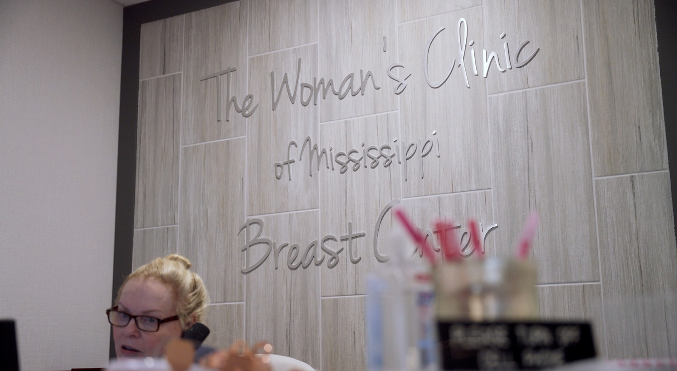
Our breast imaging center has been part of how The Woman's Clinic cares for women since well before advanced screening became standard. We are independently owned and rooted in Jackson, and we were the first private OB/GYN office in the Jackson metro area to offer Hologic 3D mammograms. Screening is not a side service here. It is central to catching cancer early, when treatment works best.
The radiologist who reads your mammogram
Here is the difference that does not show up in a brochure. At most imaging centers, your mammogram is read by a general radiologist who also reads chest X-rays, abdominal scans, and dozens of other image types on the same shift.

At The Woman's Clinic, your images go to
Dr. Susan Shamburger.
Dr. Shamburger is fellowship-trained in breast imaging and board-certified, with 18 years of experience. She previously served as associate professor of radiology and chief of breast imaging at the University of Mississippi Medical Center.
When she tells you a scan is clear, or that you need a closer look, that judgment comes from a career built on this one area of medicine. You can learn more about Dr. Susan Shamburger, fellowship-trained breast radiologist and her approach to reading your images.
Breast imaging services
Our breast imaging services
The Breast Center brings screening, diagnosis, and expert interpretation together in one place. Our core services work as a connected system, not a scattered set of appointments.

Clinical breast exams
A hands-on exam by your provider remains an important part of screening. It works alongside imaging to catch changes that deserve a closer look, and it is often where your screening conversation starts.

2D and 3D mammography
We use the Hologic 3Dimensions system for both standard and 3D mammography. A Hologic 3D mammogram captures your breast tissue in thin layers rather than a single flat image, which helps our radiologist see through overlapping tissue and spot small cancers earlier. Explore our 3D and 2D mammography to understand what each involves.

ABUS for dense tissue
For women with dense breasts, we offer the Invenia Automated Breast Ultrasound System, known as ABUS. It supplements a mammogram with ultrasound imaging designed specifically for dense tissue, where cancers are harder to detect on a mammogram alone. Learn more about ABUS screening and whether it fits your needs.
Know your risk
Screening is not one-size-fits-all, and your personal risk shapes when you should start and how often you should come in. Some risk factors you cannot change, and some you can influence. Either way, knowing yours turns screening from a routine box to check into a plan built around you.
Several factors can raise breast cancer risk:
Family and personal history
A mother, sister, or daughter with breast cancer raises your risk, as does a prior breast cancer diagnosis of your own. Certain inherited gene changes, like BRCA1 and BRCA2, raise it significantly.
Age
Risk climbs as you get older, which is why consistent screening matters more with each passing decade.
Dense breast tissue
As covered above, dense tissue both raises risk and makes cancer harder to spot on a standard mammogram.
Reproductive and hormonal factors
Starting your period early, reaching menopause late, or never having been pregnant can each play a role.
Lifestyle factors
Alcohol use, excess weight after menopause, and inactivity are among the risks you have some power to change.
Higher risk does not mean a diagnosis is coming. It means you and your provider can be more deliberate: starting screening earlier, screening more often, or adding supplemental imaging like ABUS. Bring your family history to your next visit, and we will build the right screening plan together.

Dense breast tissue
What dense breast tissue means for your screening
Dense breast tissue is common, and most women who have it never knew until a mammogram report mentioned it. Being dense simply means you have more glandular and connective tissue than fatty tissue. It is normal, and it is not something you did or can change. But it matters for two reasons, and no one should let it slide past you unexplained.
First, dense tissue raises your risk of breast cancer somewhat on its own.
Second, and just as important, dense tissue shows up white on a mammogram, and so do tumors. That makes cancers harder to see, like trying to spot a snowflake against a white sheet.
This is exactly where ABUS earns its place. By adding ultrasound built for dense tissue, we give our radiologist a clearer picture and give you a more complete screen. If your reports have ever mentioned density, read what you should know about dense breast tissue so you understand your own results.
Screening built around your comfort
A mammogram carries real anxiety for a lot of women, and a rushed, cold experience only makes it worse. We have built ours to feel different. Our team explains each step, gives you time, and treats the appointment as a conversation rather than a conveyor belt. You are a woman being cared for, not a scan on a schedule.
That care extends to what happens next. When a result needs follow-up, you are not left refreshing a portal or waiting on a callback from a stranger. Your provider and our radiologist are part of the same practice, so the path from screening to answer stays clear and quick.
Know what to watch for
Screening does the heavy lifting, but knowing your own body matters too. Most breast cancers are found through imaging, yet some are first noticed by women themselves. It helps to know what a concerning change can feel like, and that not every warning sign is a lump. Learn about recognizing malignant breast lumps and some less common signs of breast cancer so you know when to call.
And if you have been putting off this year's mammogram, you are not alone, but it is worth reconsidering. See why you shouldn't skip your mammogram, straight from Dr. Shamburger.
Visit the Breast Center
The Breast Center operates from its own dedicated suite at 501 Marshall Street, Suite 401 in Jackson, separate from the main OB/GYN clinic in Suite 400.
Screening is available at this Jackson location.
You can also meet our providers to see the full team behind your care, and find directions through our Jackson office.
When was your last mammogram read by someone who only reads mammograms?
Schedule your screening online, or call 601-354-0995.
Frequently Asked Questions
A 2D mammogram takes flat images of the breast from two angles. A 3D mammogram, or tomosynthesis, captures many thin layers, letting the radiologist scroll through the tissue rather than viewing it all stacked in one image. This makes it easier to detect small cancers and reduces false alarms, especially in dense tissue.
ABUS stands for Automated Breast Ultrasound System. It uses ultrasound to image the whole breast and is designed for women with dense breast tissue, where mammograms are less able to detect cancer. It supplements a mammogram rather than replacing it. If your reports mention dense breasts, ask whether ABUS is right for you.
Dense breast tissue means you have more glandular and connective tissue than fatty tissue. It is common and completely normal. It matters for screening because dense tissue can hide cancers on a standard mammogram and slightly raises breast cancer risk. Women with dense breasts often benefit from supplemental screening like ABUS.
Most women benefit from a screening mammogram every year starting at age 40, though your ideal schedule depends on your personal and family history. Women at higher risk may start earlier or screen more often. Your provider will recommend the right timing for you based on your individual risk.
Many insurance plans now cover 3D mammography, and coverage has expanded significantly in recent years. Plans differ, though, and some may apply it differently than standard screening. Before your appointment, call our Breast Center at 601-354-0995 and we will help you understand how your specific plan handles 3D imaging.
During a breast ultrasound, including ABUS, a technologist applies gel and moves a scanner across the breast while sound waves create detailed images. It is painless, uses no radiation, and typically takes only a short time. The images then go to our fellowship-trained radiologist for interpretation.
At The Woman's Clinic Breast Center, your mammogram is read by Dr. Susan Shamburger, a fellowship-trained, board-certified breast radiologist who reads breast imaging exclusively. Because she works within the same practice as your provider, results and any follow-up stay coordinated rather than passed between separate systems.